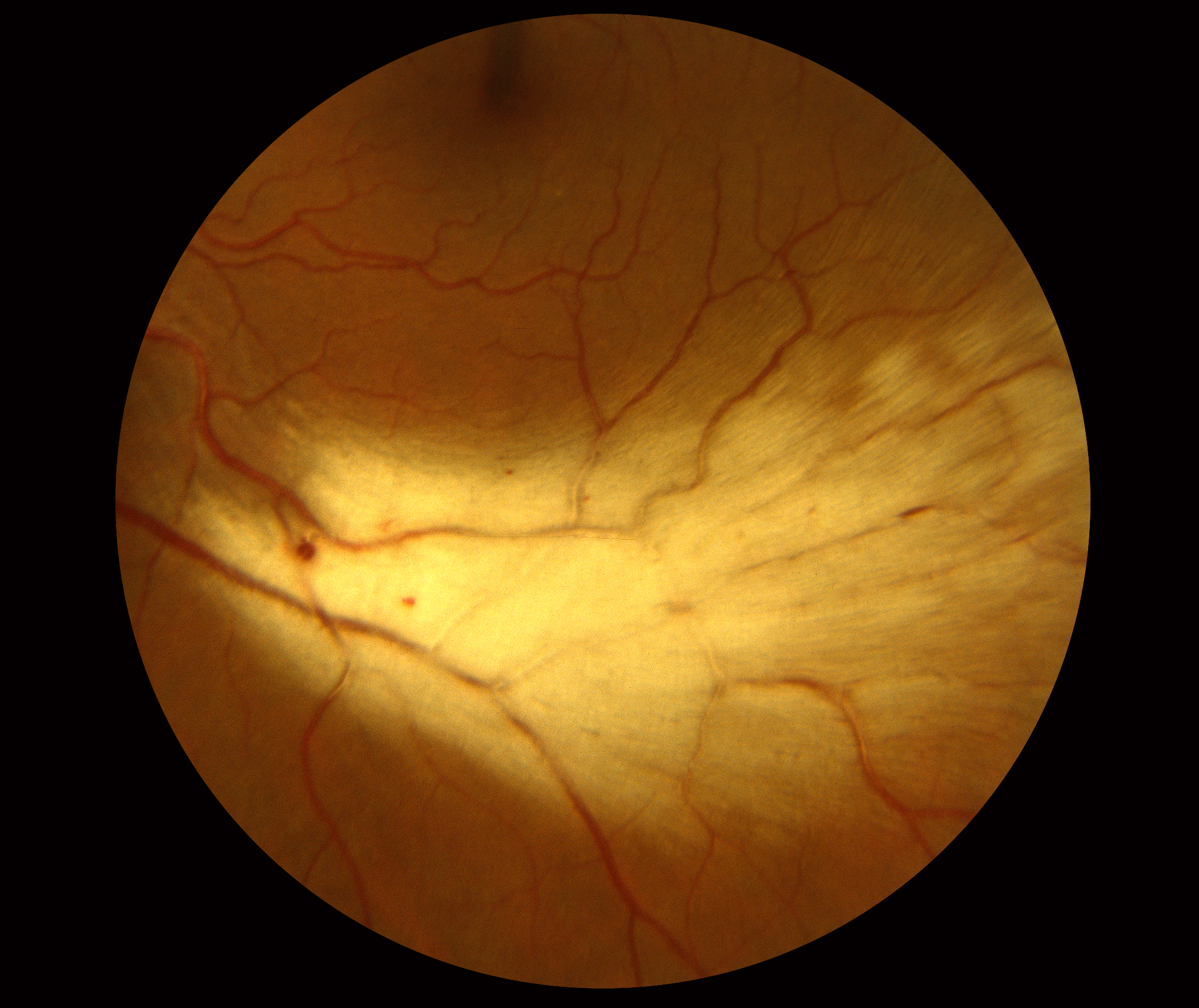

The morning glory disc anomaly is a congenital, typically unilateral funnel-shaped excavation of the posterior fundus that incorporates the optic disc, causing the disc to be markedly enlarged and appear elevated in some patients. Epipapillary glial tissue may be associated with Bergmeister’s papillae and produce anterior traction that elevates the disc. Bergmeister’s papillae is a congenital variant of the optic nerve in which an incompletely regressed hyaloid artery can obscure the optic nerve and give the impression of papilledema. In tilted optic discs, a typically bilateral condition, the superotemporal optic disc is elevated, mimicking optic disc swelling, while the inferonasal disc is posteriorly displaced. Hypoplastic optic discs are common and may be anomously elevated. The presence of myelin may elevate adjacent portions of the optic disc and obscure the disc margin and underlying retinal vessels, simulating papilledema. Myelinated nerve fibers refers to the atypical myelination of intraocular nerve fibers, occuring in 0.3-0.6% of the population by ophthalmoscopy. Tilted disc syndrome Congenital anomaliesĬongenital anomalies of the optic disc may also mimic papilledema by causing portions of the disc to elevate. For example, disc drusen may be associated with retinitis pigmentosa, Usher syndrome, pseudoxanthoma elasticum, angioid streaks, Noonan syndrome, Alagille syndrome, megalencephaly, and migraine headaches. Optic nerve head drusen can be inherited as part of a genetic syndrome with other ocular or systemic manifestations. Deeper, “buried” drusen are not visible with fundoscopy and maybe difficult to differentiate from papilledema. Over time, the drusen can result in elevation of the optic nerve head. Drusen consists of extracellular deposits of calcium, hyaline, and other proteins caused by impaired axoplasmic flow resulting in extrusion of drusen or degeneration of the retinal ganglion cell nerve fiber axons. A complete review of optic disc drusen can be found on the optic disc drusen page. Optic nerve head drusen is the most common cause of pseudopapilledema, occurring in 0.34%-2.4% of individuals. Optic nerve drusen on CT scan of the orbit Optic nerve head drusen Pseudopapilledema can be caused by a number of local and systemic processes.
#Myelinated retinal nerve fiber layer how to
In this article, we will cover multiple causes of pseudopapilledema and how to differentiate them from true papilledema. Patients thought to have papilledema are often subjected to lumbar puncture, MRI, and extensive laboratory studies to find the underlying cause. It is important to distinguish pseudopapilledema from true papilledema, which can be the first sign of disease process with the potential for vision loss, neurological impairment, or death. Papilledema, on the other hand, is a swelling of the optic disc due to increased intracranial pressure. Pseudopapilledema is defined as anomalous elevation of one or both optic discs without edema of the retinal nerve fiber layer. 2.3.6 Magnetic resonance imaging (MRI) and Magnetic resonance venography (MRV).2.3.3 Optical Coherence Tomography Angiography (OCTA).2.3.2 Optical coherence tomography (OCT).2.3.1 A-scan and B-scan ultrasonography.


 0 kommentar(er)
